



#FEMSmicroBlog: New tools to investigate DNA-binding proteins in the model archaeon Haloferax volcanii
DNA-binding proteins are essential for life as they are involved in crucial processes like replication, transcription, and DNA repair. To better understand their mechanistic functions, dynamics, and spatiotemporal pattern, super-resolution microscopy techniques with single-molecule resolution are needed. The article “A novel expression system for imaging single-molecule fluorescence in Haloferax volcanii WR806 enables visualization of altered Cas1 dynamics during UV-induced DNA damage response”, part of the Thematic Issue “CRISPR-CAS” in microLife, established an imaging toolbox for this model archaeon. In this #FEMSmicroBlog, Paula Schrage explains how these tools can help us understand time- and dosage-dependent responses to UV-light-induced DNA damage. #FascinatingMicrobes
DNA-binding proteins are essential prerequisites for life
Haloferax volcanii is one of the best-established model organisms within archaea, the third domain of life. Originally isolated from the Dead Sea, it requires high salinity to grow, is naturally exposed to elevated UV radiation, and carries an unusually high number of genome copies per cell — features that make it a compelling system for studying DNA maintenance and repair mechanisms.
Across all domains of life, DNA-binding proteins physically interact with DNA and help coordinate essential cellular processes, such as replication, transcription, and DNA repair. Understanding their molecular mechanisms requires tools that resolve molecules individually, with both high spatial precision and temporal resolution.
Super-resolution fluorescence microscopy and, particularly, single-particle tracking provide exactly these functions. Researchers can follow the dynamics of individual proteins inside living cells with nanometer-scale precision and in real time.
A new platform for single-molecule imaging in Haloferax volcanii
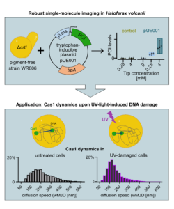
To achieve single-molecule resolution, imaging routines often have to be adapted to the biological conditions of different organisms. In Haloferax volcanii, single-molecule resolution depends on two critical prerequisites: 1) a minimal cellular background to ensure single-molecule detection and 2) precise, titratable control over protein expression levels.
The study “A novel expression system for imaging single-molecule fluorescence in Haloferax volcanii WR806 enables visualization of altered Cas1 dynamics during UV-induced DNA damage response” in microLife addressed both requirements by combining an imaging strain with a newly engineered expression system.
The strain WR806 lacks the red pigment bacterioruberin which is natively localized in the membrane. Its absence reduces background noise, enabling the visualization of single, fluorescently labelled molecules.
For controlled protein expression, they developed pUE001, an expression plasmid for WR806. In previous plasmids, the amino acid tryptophan served both as selection compound and inducer triggering protein production. Carrying the tryptophan synthetase gene, pUE001 decouples selection from induction. Protein production can be precisely and continuously adjusted by varying the supply of external tryptophan, providing a titratable expression range that was previously inaccessible.
Cas1 responds to UV-damage
With this platform established, the researchers investigated Cas1, a natively low-abundant protein that is part of the Type I-B CRISPR-Cas system of Haloferax volcanii. While CRISPR-Cas systems are best known for their role in adaptive immunity against foreign genetic elements, e.g. phages, Cas1 can also be involved in DNA repair.
Using the newly established pUE001, they expressed a fluorescently tagged version of Cas1 at low levels. They then exposed the cells to different doses of UV-light and monitored Cas1 dynamics.
Cas1 mobility slowed down significantly, in a dose- and time-dependent manner. This slow down went along with more frequent Cas 1 engagement. Even though not explicitly shown, Cas 1 then likely interacts with sites encoding or regulating the nucleotide excision repair machinery, the primary mechanism for repairing UV-light-induced lesions.
The most pronounced response was observed three hours after exposure to a medium UV dose. Higher doses produced a more complex pattern, likely reflecting the involvement of additional repair pathways or the onset of growth arrest upon severe damage levels.
This work established an imaging platform for Haloferax volcanii, consisting of a low-autofluorescence strain with a titratable expression plasmid. The system is now available for studying proteins at single-molecule resolution and provides the first in vivo imaging evidence of Cas1 dynamically responding to UV-induced DNA damage, complementing and extending earlier biochemical data.
Together, these results reinforce that DNA repair, replication, and immune defense are deeply interconnected processes. As shown here, single-molecule imaging in archaea can be a powerful tool for disentangling them.
- Read the article “A novel expression system for imaging single-molecule fluorescence in Haloferax volcanii WR806 enables visualization of altered Cas1 dynamics during UV-induced DNA damage response” in the Thematic Issue “CRISPR-CAS” by Schrage et al. in microLife (2026).
 Paula Schrage is a doctoral student at the research group of Prof. Ulrike Endesfelder at the University of Bonn where she investigates archaeal cell biology of H. volcanii. After finishing her master’s in microbiology, she decided to continue her education in the interdisciplinary research field of super-resolution microscopy in microbes. She is particularly interested in the detailed and mechanistic understanding of the dynamics and processes of the essential and evolutionarily important DNA-binding proteins in the halophile H. volcanii using both classical microbiological and biochemical tools as well as state-of-the art single-molecule sensitive techniques.
About this blog section
The section #FascinatingMicrobes for the #FEMSmicroBlog explains the science behind a paper and highlights the significance and broader context of a recent finding. One of the main goals is to share the fascinating spectrum of microbes across all fields of microbiology.
| Do you want to be a guest contributor? |
| The #FEMSmicroBlog welcomes external bloggers, writers and SciComm enthusiasts. Get in touch if you want to share your idea for a blog entry with us! |