


Understanding the cellular architecture of microbes can provide better insights into their physiology. The study “Phylogenetic diversity of core rumen microbiota as described by cryo-ET” in microLife uses advanced microscopy techniques to get a detailed snapshot into the microbial world. In a #BehindThePaper interview, Itzhak Mizrahi, Sarah Morais, Benedikt Wimmer and Ohad Medalia explain how these results help us better understand microbial phylogeny and evolution.
Can you summarise the significance of your paper for microbiologists outside of your field?
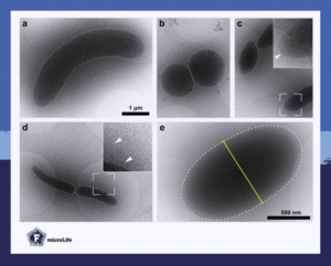
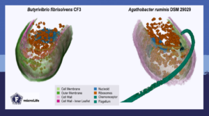
Our study provides a new scientific perspective on microbial ecology and evolution. We examined microbes and their cellular architecture with cryo-electron microscopy to get a more comprehensive understanding of their diversity and behavior.
Traditionally, the microbes’ perspective has been neglected, so that we could barely understand their behavior and the dynamics of microbial communities. However, with the advancement of cryo-electron microscopy, we can now capture the microscopic view of microbes and gain insights into their world.
With cryo-electron microscopy, we created an atlas of bacterial and archaeal species of rumen origin. Now, we have archive of 2D electron micrographs of about 70 strains and 3D electron tomograms of 40 strains. By comparing the cellular architecture of these strains along five dimensions, we found that closely related species do indeed look very similar.
What can policymakers learn from your research results?
Our results highlight the importance of considering cellular architecture when assessing the relationships between microbes. By recognizing the significance of the intricate details of microbial architecture and behavior, policy makers can gain a more comprehensive understanding of microbial diversity and behavior in ecological systems.
Our study serves as an initial step to use cellular architecture as a tool for studying microbial ecology and ecosystems. In the future, we hope that structural biology and ecology will be considered together. Ultimately, our research can help inform policies that promote sustainable microbial management and contribute to the preservation of ecological systems.
Can you explain the relevance of your article to non-scientists?
Our article sheds light on the microscopic world of microbes and their importance in maintaining healthy ecosystems. It provides a new perspective on the behavior and diversity of microbes, which can ultimately lead to the development of new approaches for engineering microbial communities for environmental and health-related applications.
Why did you choose to dive into the topic of this paper? What fascinates you about cellular morphologies?
Studying the life of microbes at the micro and nanoscale is an incredibly exciting and promising area of research. The unique properties and behaviors of microorganisms at this level of detail are incredibly fascinating. We are intrigued to gain a deeper understanding of how these tiny organisms interact with their environment, other organisms and each other.
While studying the structure of microorganisms falls under the realm of structural biology, we were fascinated to recognize microbial structures are closely related to their phylogeny and ecological interactions. We hope that researchers will consider these two fields more and more complementary.
By using advanced microscopy technologies such as cryo-electron microscopy, we are now unlocking more secrets of microbes and learning how their lives unfold. Applying both structural biology and ecology together provides valuable insights into the natural world and potentially to develop new approaches to preserving and protecting ecosystems.
In summary, the complexity of microbial interactions and behaviors at the micro and nanoscale is what fascinates us the most about this topic. Plus, we believe that studying these tiny organisms will lead to new insights into the natural world.
- Read the paper “Phylogenetic diversity of core rumen microbiota as described by cryo-ET” by Wimmer et al. (2023).
About the authors of this blog
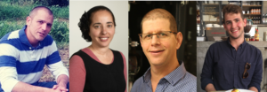
Itzhak Mizrahi is a professor at BGU, where he leads the microbial ecogenomics group. His research focuses on the microbial ecology of the gut microbiome of vertebrate animals, with a particular interest in ruminant microbiomes. He explores fundamental basic questions related to microbial community assembly, as well as issues of food security and agricultural sustainability, aligning with the One Health approach. Through his research, Prof. Mizrahi seeks to make a significant impact on our understanding of the complex relationship between animals, their gut microbiomes, and the environment.
Dr Sarah Morais is an internationally recognized scientist in the field of plant fiber degradation. She is acting as a staff scientist in Prof. Itzhak Mizrahi microbial ecogenomic group in Ben Gurion University in Israel. She applies her solid expertise in plant fiber degradation biochemistry and microbiology to explore gut microbial ecosystems ecology and function.
Benedikt Wimmer is a PhD student in Ohad Medalia’s group at the Department of Biochemistry, University of Zurich. His research focuses on applying advanced 3D electron microscopy to study bacterium–substrate interactions in microbiomes.
Ohad Medalia is an acknowledged expert in the field of cryo-electron tomography. The research of his group focuses on macromolecular structures and their interactions, mostly in eukaryotic cells. In particular, Prof. Medalia is interested in cytoskeletal alterations that are associated with the mechanical stress of cells. This includes cell adhesion, intermediate filament assemblies, and the organization of the nuclear envelope. In recent years, the group also studies the mystery of the plant fiber degradation machineries, using innovative structural approaches.
About this blog section
#BehindThePaper posts on the #FEMSmicroBlog aim to bring science closer to different audiences and to tell more about the scientific or personal journey to come to the results.
| Do you want to be a guest contributor? |
| The #FEMSmicroBlog welcomes external bloggers, writers and SciComm enthusiasts. Get in touch if you want to share your idea for a blog entry with us! |